Electro- and Magnetoneurography
 |
||
| Contents: | ||
More than 6 % of the population suffers from peripheral neuropathies. Over 90 % of all neuropathies are induced by local pressure on the nerve. A detailed diagnosis requires exact localization and treatment by conservative or surgical decompression e.g. 300 000 surgeries are only performed for the carpal tunnel syndrome every year.
 Magnetoneurography (MNG) provides an alternative and also very attractive technology to the electrode-based nerve conduction studies called Electroneurography (ENG). The Electroneuography is the current gold standard of nerve assessment to investigate the nerve conduction velocity. In this case the nerve signal is deviated by conventional electrical electrodes. This technique only allows to distinguish between axonal and demyelinating forms of neuropathy, but the localizing capacities are quite poor. Finally, this results in a lack of spatial resolution, because only segment-wise information are available. Additional modern nerve imaging techniques are required to create a functional projection of the nerve characterizing for an accurate diagnosis and treatment. Hence, our first objective is to optimize the diagnostic specificity via a multichannel neurographical approach and to apply new signal processing methods for further signal enhancement.
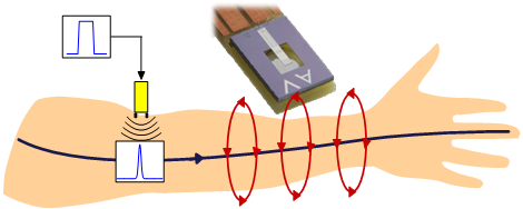
Magnetoneurography uses magnetic field sensors instead and they do not require direct skin contact. Moreover magnetic field sensors can be combined in arrays to make a localization of the potential nerve pathology possible. This enables also highly resolved functional nerve sampling at bedside. The main objective of this modern technique is to optimize the diagnostic specificity of neuropathies by developing a spatially continuous scanning of nerves with a magnetic field sensor. This approach will increase and also combine the spatial resolution and functional information. In summary, this will further improve the treatment.
The nerve electromagnetic field of human nerve pulses is quite low. Peak amplitudes in the range of ft to pT can be achieved by using an external electric stimulation of the nerve. As the stimulus to the nerve can be repeated, the signal to noise ratio can be improved by using enhanced low-noise adaptive averaging algorithms. Project B6 of the Collaborative Research Centre CRC 1261 focuses on the Electro- and Magnetoneurography and also the signal to noise enhancement of such nerve signals. This project founded by the Deutsche Forschungsgemeinschaft uses new sensor concepts to realize a Magnetoneurography at room temperature. Further details about this project of the CRC 1261 can be found here.
Corresponding Publications:
E. Elzenheimer, H. Laufs, W. Schulte-Mattler, G. Schmidt: Magnetic Measurement of Electrically Evoked Muscle Responses with Optically Pumped Magnetometers, IEEE Transactions on Neural Systems and Rehabilitation Engineering, January 2020, doi: 10.1109/TNSRE.2020.2968148
E. Elzenheimer, H. Laufs, W. Schulte-Mattler, G. Schmidt: Signal Modeling and Simulation of Temporal Dispersion and Conduction Block in Motor Nerves, IEEE Transactions on Biomedical Engineering, November 2019, doi: 10.1109/TBME.2019.2954592
E. Elzenheimer, H. Laufs, W. Schulte-Mattler, G. Schmidt: Magnetic Measurement of Electrically Evoked Muscle Responses with Optically Pumped Magnetometers (OPMs), New opportunities in biomagnetism using OPMs, Biomedical Engineering / Biomedizinische Technik, 64, 39 (2019), doi: 10.1515/bmt-2019-6008
E. Elzenheimer, H. Laufs, T. Sander-Thömmes, G. Schmidt: Magnetoneurograhy of an Electrically Stimulated Arm Nerve, Joint Journal of the German Society for Biomedical Engineering in VDE and the Austrian and Swiss Societies for Biomedical Engineering and the German Society of Biomaterials, Volume 63, Number 12, Pages 363-366, September 2018
E. Elzenheimer, F. Weitkamp, H. Laufs, T. Sander-Thömmes, G. Schmidt: Magnetoneurografie eines elektrisch stimulierten Armnervs, Biosignale Workshop, 2018, Erfurt, Germany
